43 labelling of compound microscope
nationalpost.com › category › newsLatest Breaking News, Headlines & Updates | National Post Read latest breaking news, updates, and headlines. Get information on latest national and international events & more. Lifestyle | Daily Life | News | The Sydney Morning Herald WebThe latest Lifestyle | Daily Life news, tips, opinion and advice from The Sydney Morning Herald covering life and relationships, beauty, fashion, health & wellbeing
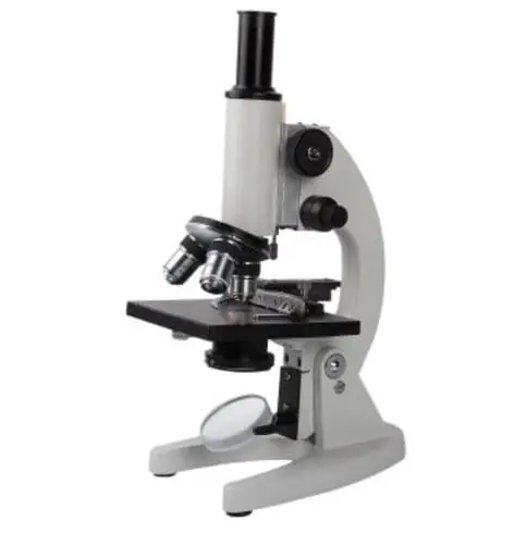
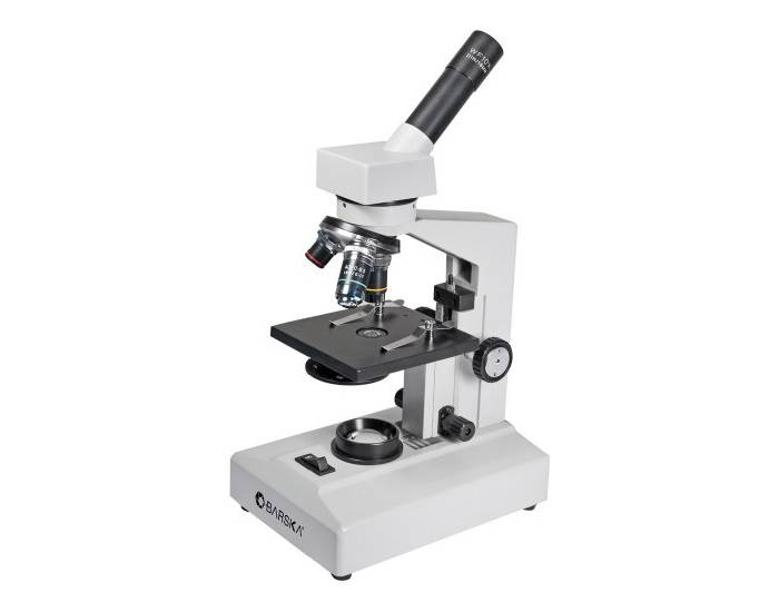
A Study of the Microscope and its Functions With a Labeled Diagram The compound microscope uses light for illumination. Some compound microscopes make use of natural light, whereas others have an illuminator attached to the base. The specimen is placed on the stage and observed through different lenses of the microscope, which have varying magnification powers. Compound Microscope Parts and Functions

Labelling of compound microscope
label parts of a compound microscope - TeachersPayTeachers This is a set of 3 tiered readings. Students will read a passage about the how to use a compound light microscope. Students will use textual evidence to answer questions and label the different parts of the microscope. It also allows students to gain prior knowledge about the compound microscope. Version A provides the most support for students. Labeling the Parts of the Microscope | Microscope activity, Science ... More like this. Bioimager is a professional microscopy company to provide custom microscopy solution for life science and industrial applications at great price & quality. Compound microscope is a microscope that uses multiple lenses to enlarge the image of sample. *This post contains affiliate links. Parts of a syringe (plain tip) and needle. What is a Compound Microscope? - New York Microscope Company A compound microscope is an instrument that is used to view magnified images of small specimens on a glass slide. It can achieve higher levels of magnification than stereo or other low power microscopes and reduce chromatic aberration. It achieves this through the use of two or more lenses in the objective and the eyepiece.
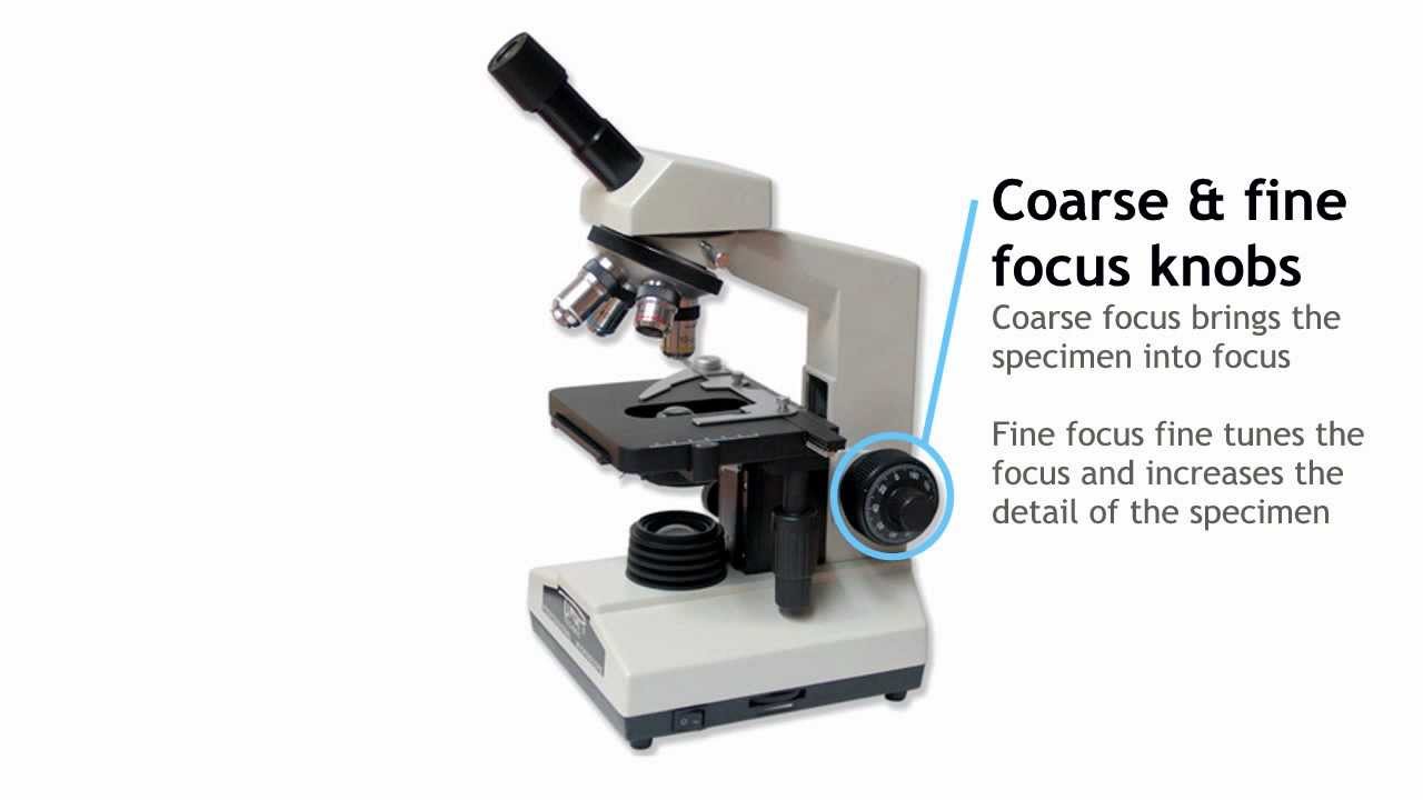
Labelling of compound microscope. Electron microscope - Wikipedia WebAn electron microscope is a microscope that uses a beam of accelerated electrons as a source of illumination. As the wavelength of an electron can be up to 100,000 times shorter than that of visible light photons, electron microscopes have a higher resolving power than light microscopes and can reveal the structure of smaller objects. A scanning … Latest Breaking News, Headlines & Updates | National Post WebRead latest breaking news, updates, and headlines. Get information on latest national and international events & more. › articles › s41586/018/0287-8Targeting STING with covalent small-molecule inhibitors | Nature Jul 04, 2018 · a, Labelling of endogenous STING immunoprecipitaed from splenocytes of mice treated with C-176-AL, visualized by in-gel fluorescence. One representative of n = 2. Parts of the Microscope with Labeling (also Free Printouts) A microscope is one of the invaluable tools in the laboratory setting. It is used to observe things that cannot be seen by the naked eye. Table of Contents 1. Eyepiece 2. Body tube/Head 3. Turret/Nose piece 4. Objective lenses 5. Knobs (fine and coarse) 6. Stage and stage clips 7. Aperture 9. Condenser 10. Condenser focus knob 11. Iris diaphragm
Compound Microscope- Definition, Labeled Diagram, Principle, Parts, Uses A compound microscope is of great use in pathology labs so as to identify diseases. Various crime cases are detected and solved by drawing out human cells and examining them under the microscope in forensic laboratories. The presence or absence of minerals and the presence of metals can be identified using compound microscopes. Compound Microscope Parts The three basic, structural components of a compound microscope are the head, base and arm. Head/Body houses the optical parts in the upper part of the microscope Base of the microscope supports the microscope and houses the illuminator Arm connects to the base and supports the microscope head. It is also used to carry the microscope. Compound Microscope: Parts of Compound Microscope - BYJUS The parts of the compound microscope can be categorized into: Mechanical parts Optical parts (A) Mechanical Parts of a Compound Microscope 1. Foot or base It is a U-shaped structure and supports the entire weight of the compound microscope. 2. Pillar It is a vertical projection. This stands by resting on the base and supports the stage. 3. Arm Microscope Labeling Game - PurposeGames.com Microscope Labeling Game by sloanescience 2,126,978 plays 15 questions ~ 40 sec 518 4.07 (you: not rated) Language English Tries Unlimited [?] Last Played December 7, 2022 - 08:01 PM There is a printable worksheet available for download here so you can take the quiz with pen and paper. Remaining 0 Correct 0 Wrong 0 Press play! 0% 0:00.0 Highscores
Compound Light Microscope: Everything You Need to Know A compound light microscope is a type of light microscope that uses a compound lens system, meaning, it operates through two sets of lenses to magnify the image of a specimen. It's an upright microscope that produces a two-dimensional image and has a higher magnification than a stereoscopic microscope. It also goes by a couple of other names ... › nature › articlesBrowse Articles | Nature Dec 09, 2022 · A method to edit the backbones of molecules allows chemists to modify ring-shaped chemical structures with greater ease. Compound Microscope Parts - Labeled Diagram and their Functions The term "compound" refers to the microscope having more than one lens. Basically, compound microscopes generate magnified images through an aligned pair of the objective lens and the ocular lens. In contrast, "simple microscopes" have only one convex lens and function more like glass magnifiers. A bioorthogonal system reveals antitumour immune function of Web11.03.2020 · a, NP–GFP was treated with Phe-BF 3 or another indicated fluorine compound. The reactions were centrifuged (Extended Data Fig. 4b).P, pellet; S, supernatant; T, total;*, loading control sampled ...
Compound Microscope Parts and Functions | Microscope parts ...
Join LiveJournal WebPassword requirements: 6 to 30 characters long; ASCII characters only (characters found on a standard US keyboard); must contain at least 4 different symbols;

Compound Microscope Parts, Functions, and Labeled Diagram ...
Compound Microscope: Definition, Diagram, Parts, Uses, Working ... - BYJUS The compound microscope is mainly used for studying the structural details of cell, tissue, or sections of organs. The parts of a compound microscope can be classified into two: Non-optical parts Optical parts Non-optical parts Base The base is also known as the foot which is either U or horseshoe-shaped.

Compound Microscope: Parts of Compound Microscope
What is a Compound Microscope? | Microscope World Blog A compound microscope is an upright microscope that uses two sets of lenses (a compound lens system) to obtain higher magnification than a stereo microscope. A compound microscope provides a two-dimensional image, while a stereo microscope provides a three-dimensional image. Compound microscopes typically provide magnification in the range of ...

Can someone can send me diagram of this compound microscope ...
pubchem.ncbi.nlm.nih.gov › compound › 2_4-Di-tert2,4-Di-tert-butylphenol | C14H22O - PubChem /ALTERNATIVE and IN VITRO TESTS/ The volatile organic compound 2,4-di-tert-butyl phenol (2,4 DTBP) was purified from the cell free supernatant of a newly isolated Lactococcus sp. by solvent extraction and chromatographic techniques. Molecular characterization of the compound by ESI-MS, 1H NMR and FTIR analysis revealed the structure, C14H22O.

AmScope B400AB Compound Binocular Microscope, WF10x, WF16x ...
2,4-Di-tert-butylphenol | C14H22O - PubChem WebThe compound was detected not quantified in sediments from the River Po in the vicinity of River Ticino, River Olona and River Lambro, Milan, Italy. Sampling was conducted in winter and summer, 2005(2). (1) Jungclaus GA et al; Environ Sci Technol 12: 88-96 (1978) (2) Vigano L et al; Chemosphere 73: 1078-7089 (2008) Hazardous Substances Data Bank …
Parts of a Compound Light Microscope
Diagram of a Compound Microscope - Biology Discussion The size of objects viewed under the compound microscope can be accurately determined using a micrometer. The latter consists of two scales, the eyepiece scale, (also called 'graticule' or 'ocular') and the stage micrometer scale. The eyepiece scale is calibrated with the help of stage micrometer and the former is then used for measurements.
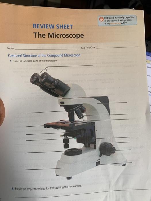
Solved Instructors may assign a portion of the Review Sheet ...
microscopeinternational.com › fluorescence-microscopyExplanation and Labelled Images - New York Microscope Company Dec 16, 2020 · The Characteristics of a Fluorescence Microscope. The main parts of a fluorescent microscope overlap with the traditional light microscope. However, there are two main features that sets fluorescent microscope apart from the traditional microscope. One is the type of light source and the other is the use of specialized filter elements.

Label the parts of a Compound Microscope.docx - A. 1. Label ...
Microscope Parts and Functions Most specimens are mounted on slides, flat rectangles of thin glass. The specimen is placed on the glass and a cover slip is placed over the specimen. This allows the slide to be easily inserted or removed from the microscope. It also allows the specimen to be labeled, transported, and stored without damage.

I. Label the parts of a Compound Microscope. You may choose ...
Labelled Diagram of Compound Microscope The below mentioned article provides a labelled diagram of compound microscope. Part # 1. The Stand: The stand is made up of a heavy foot which carries a curved inclinable limb or arm bearing the body tube. The foot is generally horse shoe-shaped structure (Fig. 2) which rests on table top or any other surface on which the microscope in kept.
Parts of a Microscope with Their Functions – Microbe Online
How to draw compound of Microscope easily - step by step I will show you " How to draw compound of microscope easily - step by step "Please watch carefully and try this okay.Thanks for watching.....#microscopedrawi...

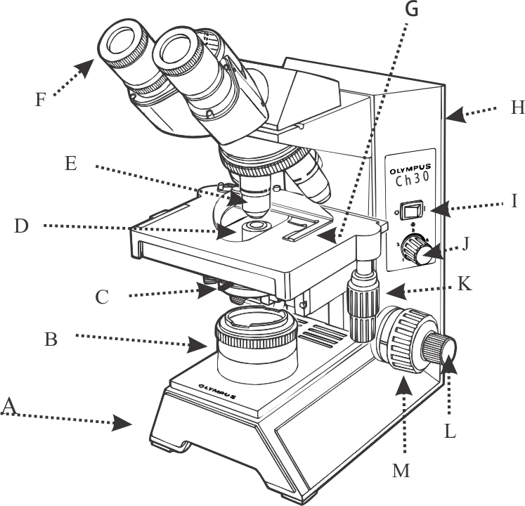
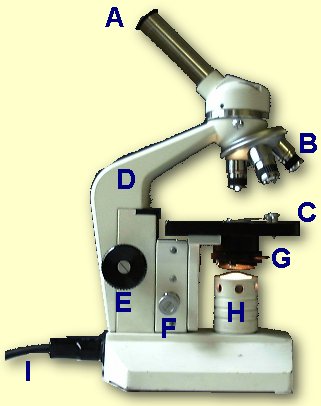
This is a common compound microscope. What the labelling D ...
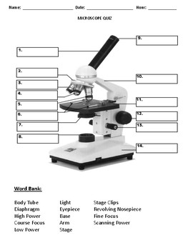
Labeling the Parts of the Microscope | Microscope World Resources Labeling the Parts of the Microscope This activity has been designed for use in homes and schools. Each microscope layout (both blank and the version with answers) are available as PDF downloads. You can view a more in-depth review of each part of the microscope here. Download the Label the Parts of the Microscope PDF printable version here.
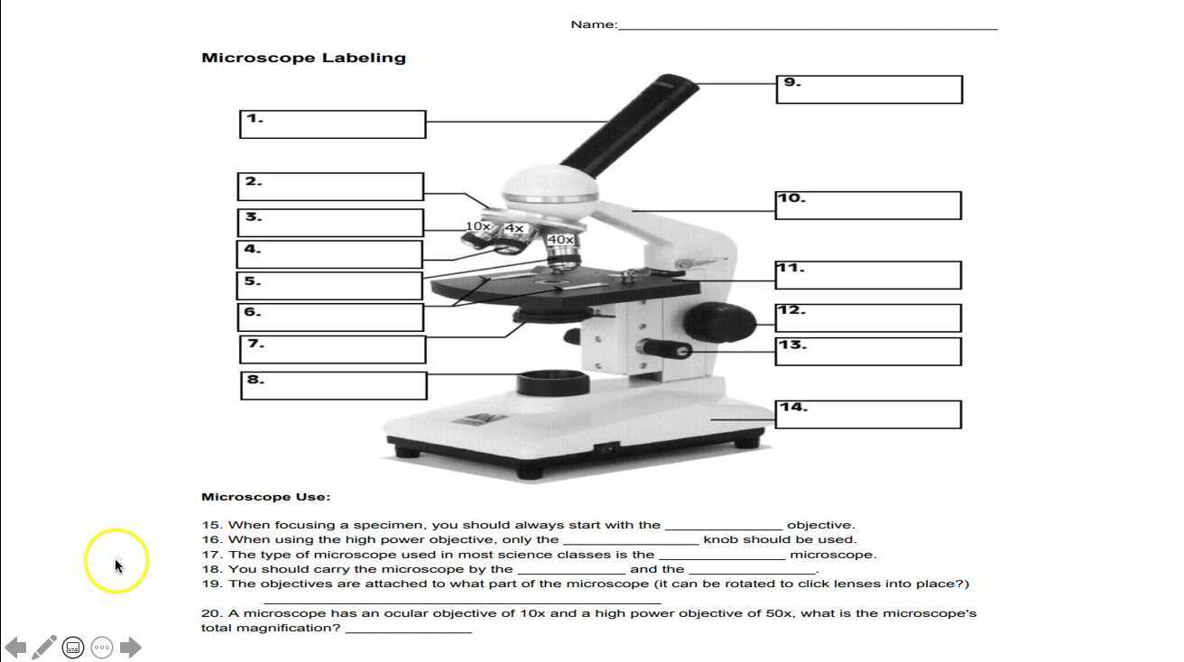
Microscope Labeling
Parts of a Compound Microscope - Labeled (with diagrams) A compound microscope is known as a high-power microscope that enables you to achieve a high level of magnification. Smaller specimens can be thoroughly viewed using a compound microscope. ... Image 3: A compound microscope with a corresponding label of the different parts. imagesource: images.slideplayer.com. The optical components of a ...

Parts of the Microscope with Labeling (also Free Printouts ...
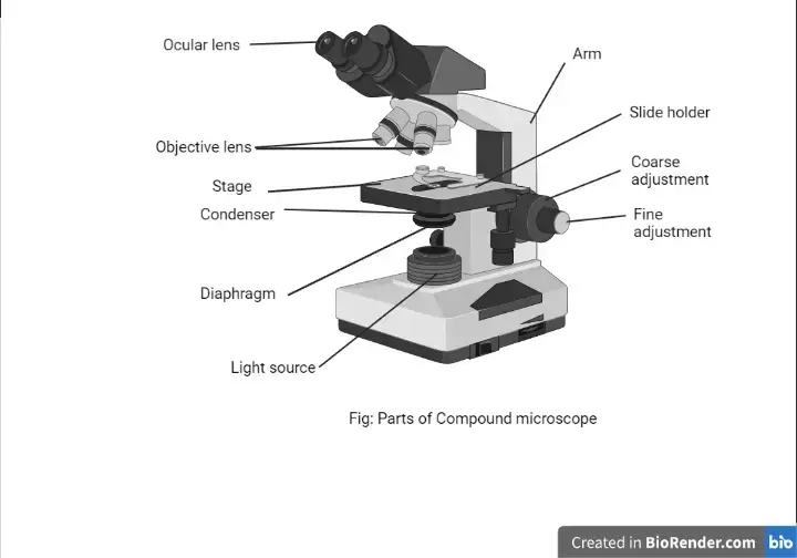
16 Parts of a Compound Microscope: Diagrams and Video The 16 core parts of a compound microscope are: Head (Body) Arm Base Eyepiece Eyepiece tube Objective lenses Revolving Nosepiece (Turret) Rack stop Coarse adjustment knobs Fine adjustment knobs Stage Stage clips Aperture Illuminator Condenser Diaphragm Video: Parts of a compound Microscope with Diagram Explained

List: Parts of a Microscope and their Function | Pathwooded
Quia - Label the Microscope Quiz Label the Microscope Quiz Choose the word that correctly labels the parts of the microscope. Please enter your name. Tools Copy this to my account E-mail to a friend Find other activities Start over Print Help Mrs. Coyle Simmons Elementary School Versailles, KY View profile This activity was created by a Quia Web subscriber. Learn more about Quia

Compound Microscope Parts, Functions, and Labeled Diagram ...
Microscope Types (with labeled diagrams) and Functions Is used to view samples that are not visible to the naked eye. Uses two types of lenses - Objective and ocular lenses. Has a higher level of magnification - Typically up to 2000x. Is used in hospitals and forensic labs by scientists, biologists and researchers to study micro organisms. Compound microscope labeled diagram.

Biology Microscope Labeling and Definitions (Light/Compound ...
Fluorescence Microscopy - Explanation and Labelled Images Web16.12.2020 · A fluorescence microscope works by combining the magnifying properties of the light microscope with fluorescence emitting properties of compounds. Fluorescence microscopy uses a high-intensity light source that excites a fluorescent molecule called a fluorophore in the sample observed. The samples are labeled with fluorophore where …
Compound Microscope - Types, Parts, Diagram, Functions and ...
Browse Articles | Nature Web09.12.2022 · A method to edit the backbones of molecules allows chemists to modify ring-shaped chemical structures with greater ease.

Compound Light Microscope Labeling Diagram | Quizlet
Parts of a Compound Microscope (And their Functions) - Scope Detective Parts of a Compound Microscope (And their Functions) By Chris Ramsay A compound microscope is the most common microscope you can get and the type you'll typically see in a lab or hobbyist's study. These microscopes tend to have total magnification between 40x - 2000x to allow you to see specimens like bacteria and cells.

Draw a neat labelled diagram of a compound microscope. Derive ...
Open Access | Open Access Publications Web» A complete version of the work and all supplemental materials, including a copy of the permission as stated above, in a suitable standard electronic format is deposited immediately upon initial publication in at least one online repository that is supported by an academic institution, scholarly society, government agency, or other well-established …
Compound Microscope. Label the numbered parts of the ...
Targeting STING with covalent small-molecule inhibitors | Nature Web04.07.2018 · Cells were collected in PBS and analysed by in-gel analysis of C-176-AL-mediated labelling of STING (see ‘ Gel-based analysis of compound binding to STING’). Immunoprecipitation

Microscope Diagram Labeled, Unlabeled and Blank | Parts of a ...
Label the microscope — Science Learning Hub Label the microscope Interactive Add to collection Use this interactive to identify and label the main parts of a microscope. Drag and drop the text labels onto the microscope diagram. eye piece lens diaphragm or iris coarse focus adjustment stage base fine focus adjustment light source high-power objective Download Exercise Tweet

Living Environment Course
en.wikipedia.org › wiki › Electron_microscopeElectron microscope - Wikipedia An electron microscope is a microscope that uses a beam of accelerated electrons as a source of illumination. As the wavelength of an electron can be up to 100,000 times shorter than that of visible light photons , electron microscopes have a higher resolving power than light microscopes and can reveal the structure of smaller objects.

Multiple Choice Quiz on Compound Microscope Parts and Functions
Compound Light Microscope Labelling Quiz - PurposeGames.com Compound Light Microscope Labelling — Quiz Information. This is an online quiz called Compound Light Microscope Labelling. There is a printable worksheet available for download here so you can take the quiz with pen and paper. Quiz Points. 15 p. You need to get 100% to score the 15 points available.

This is a common compound microscope. Label its parts from A ...
Compound Microscope Labeled Diagram | Quizlet Compound Microscope Labeled + − Flashcards Learn Test Match Created by meganplocher734 Terms in this set (14) Eyepiece/Ocular lens Contains the ocular lens Body tube A hollow cylinder that holds the eyepiece. Arm Part that supports the microscope. Stage Supports the slide or specimen Coarse adjustment Knob

Parts of a Microscope - SmartSchool Systems
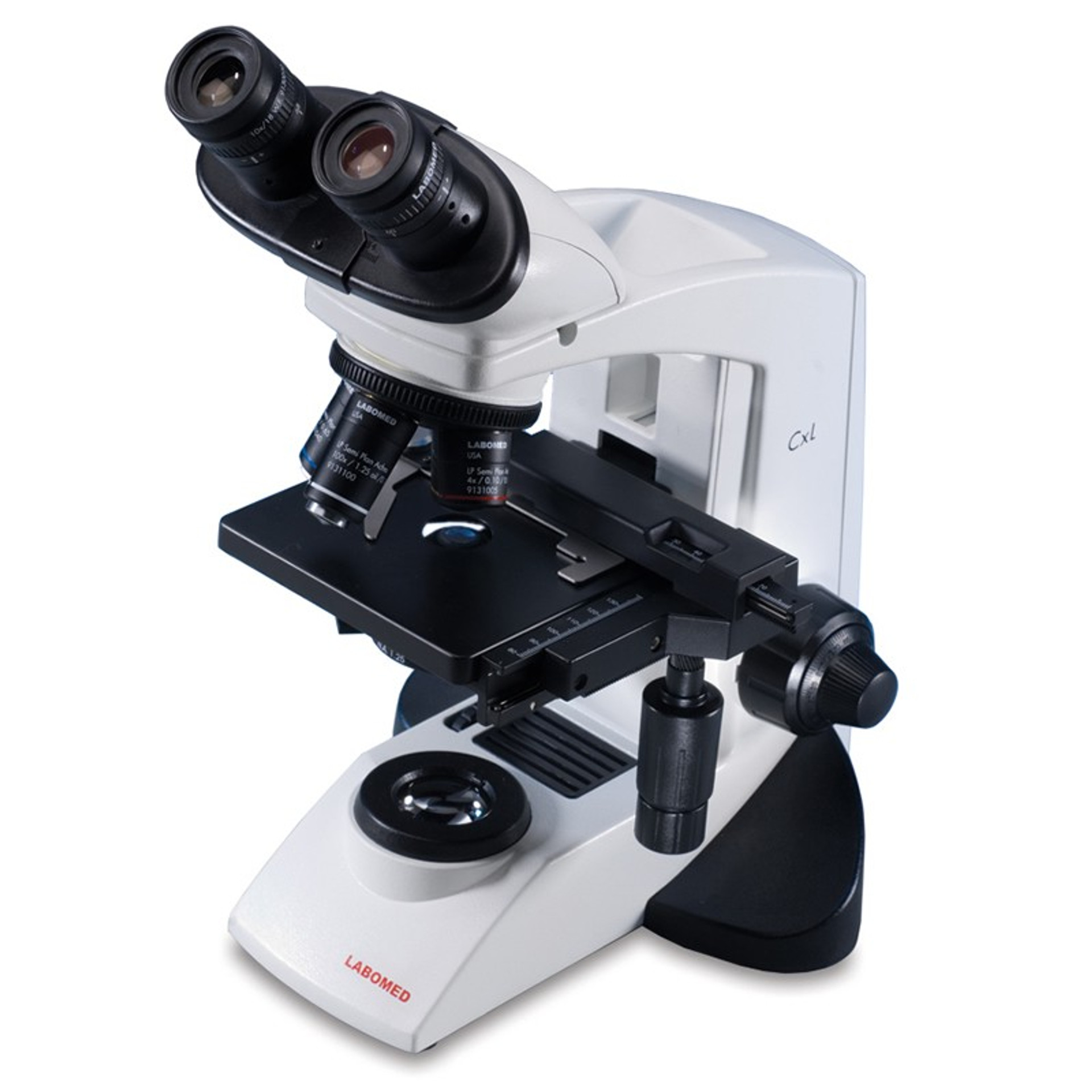
Compound Microscope Parts, Functions, and Labeled Diagram Compound Microscope Definitions for Labels Eyepiece (ocular lens) with or without Pointer: The part that is looked through at the top of the compound microscope. Eyepieces typically have a magnification between 5x & 30x. Monocular or Binocular Head: Structural support that holds & connects the eyepieces to the objective lenses.
Microscope Quiz by Quick Witted Owl Lesson Plans Made Easy | TpT
Compound Microscope - Types, Parts, Diagram, Functions and Uses Compound microscope - It is an optical instrument consists of two convex lenses of short focal lengths primarily used for observing a highly magnified image of minute objects. Lenses Simple microscope - It has a convex lens. It uses only one lens to magnify objects. An example of a simple microscope is a magnifying glass.

Biology label part of microscope
Compound Microscope - Diagram (Parts labelled), Principle and Uses Why is it called compound microscope? Because it has multiple lenses that work in conjunction to magnify a specimen Q 4. What are the 13 parts of a microscope? 1. Eyepiece 2. Eyepiece Tube 3. Objective Lens 4. Stage 5. Stage Clips 6. Nosepiece 7. Fine and Coarse Focus knobs 8. Illuminator 9. Aperture 10. Iris Diaphragm 11. Condenser 12.

easy compound microscope diagram - Clip Art Library
en.wikipedia.org › wiki › Transmission_electronTransmission electron microscopy - Wikipedia Transmission electron microscopy (TEM) is a microscopy technique in which a beam of electrons is transmitted through a specimen to form an image. The specimen is most often an ultrathin section less than 100 nm thick or a suspension on a grid.

Microscope labeling
What is a Compound Microscope? - New York Microscope Company A compound microscope is an instrument that is used to view magnified images of small specimens on a glass slide. It can achieve higher levels of magnification than stereo or other low power microscopes and reduce chromatic aberration. It achieves this through the use of two or more lenses in the objective and the eyepiece.
Using Microscopes - Bio111 Lab
Labeling the Parts of the Microscope | Microscope activity, Science ... More like this. Bioimager is a professional microscopy company to provide custom microscopy solution for life science and industrial applications at great price & quality. Compound microscope is a microscope that uses multiple lenses to enlarge the image of sample. *This post contains affiliate links. Parts of a syringe (plain tip) and needle.

Biology - labeling a compound microscope Diagram | Quizlet
label parts of a compound microscope - TeachersPayTeachers This is a set of 3 tiered readings. Students will read a passage about the how to use a compound light microscope. Students will use textual evidence to answer questions and label the different parts of the microscope. It also allows students to gain prior knowledge about the compound microscope. Version A provides the most support for students.

National 131-RLED-MS Compound Microscope 131-RLED-MS B&H Photo
Compound Microscope Parts, Functions, and Labeled Diagram ...
Label the microscope Quiz
compound-microscope-unlabeled.jpg

Microscope Components - Science Quiz
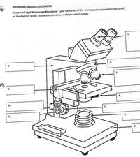
Answered: Microscope Structure and Function… | bartleby
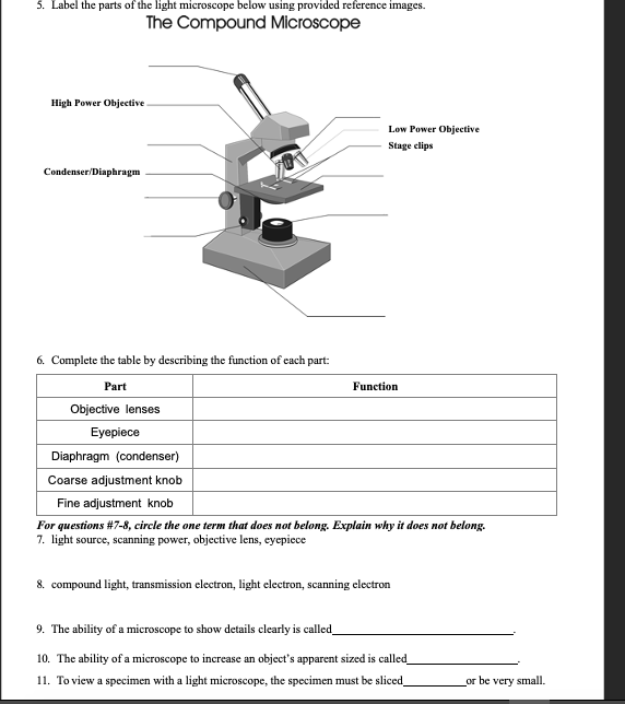
Solved 5. Label the parts of the light microscope below ...

Diagram of a Compound Microscope

Compound microscope - their parts and function - Microscopy4kids

Microscopes / Group 5
Microscope

Simple Microscope Definition, Magnification, Parts And Uses


Komentar
Posting Komentar